Types of RVO
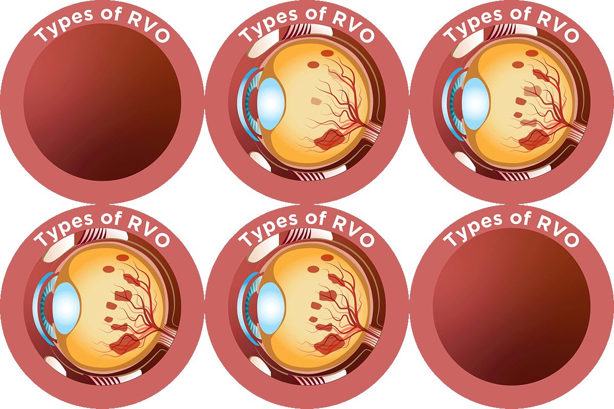
Retinal vein occlusion (RVO) can be divided into two primary categories – branch and central RVO – depending on the site of blockage (occlusion), with branch occlusions occurring more commonly than central ones.1-3
Central retinal vein occlusion (CRVO)
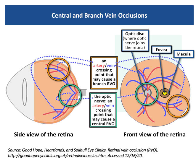
CRVO occurs when the main vein that drains blood from the retina is blocked by a blood clot or br overlying artery causing pressure on the vein, reducing blood flow either either partially or completely.3 Blockage of the vein can cause swelling in the center part of the retina, which is responsible for sharp and central (straight-ahead) vision; this is called macular edema and creates symptoms of blurry or distorted vision, typically in one eye.3,4 CRVO occurring in both eyes may be associated with diseases related to blood clotting that affect the whole body.5 CRVOs are responsible for about 20% to 30% of all retinal vein occlusions and are more likely to cause permanent vision loss than branch retinal vein occlusions.6,7
Branch retinal vein occlusion (BRVO)
BRVO is a blockage of one or more of the four smaller veins that branch off of the main central vein.5,8 BRVO usually occurs where the artery and vein cross each other; the artery wall can thicken and press on the softer vein, compressing it.8-10 Normally smooth blood flow then becomes turbulent, leading to clot formation and eventual blockage of the affected blood vessel.6,8 Just like with CRVO, the vein is not able to drain blood from the retina, which can lead to macular edema and poor blood circulation in the retina.5 Symptoms may include sudden, painless vision loss; however, no symptoms may be experienced if the area of blockage is not in the center of the eye.8
Currently, there is no treatment to remove retinal vein blockages, and the poor circulation caused by RVO can promote the growth of new, abnormal blood vessels called neovascularization, which can leak or bleed.3,4,8 Ocular Coherence Tomography (OCT) and fluorescein angiography (FA) are used to diagnose and monitor treatment progression.8 OCT is a non-invasive test that uses light waves to take pictures of the layers of the retina.4 FA involves injecting into the arm and using a special camera to take pictures of the blood vessels in the eye, which can detect growth of new, damaged vessels.4,5 Your eye care provider may repeat OCT testing to monitor for any change in retinal thickness to help guide your treatment.4

http://goodhopeeyeclinic.org.uk/retinalveinocclus.htm.
References
- Laouri M, Chen E, Looman M, Gallagher M. The Burden of disease of retinal vein occlusion: review of the literature. Eye. 2011;25:981-988. https://www.ncbi.nlm.nih.gov/pmc/articles/PMC3178209/
- The Royal College of Ophthalmologists. Clinical Guidelines. Retinal Vein Occlusion (RVO). 2022.
https://www.rcophth.ac.uk/wp-content/uploads/2015/07/Retinal-Vein-Occlusion-Guidelines-2022.pdf. - American Society of Retina Specialists (ASRS). Central retinal vein occlusion. 2020. https://www.asrs.org/patients/retinal-diseases/22/central-retinal-vein-occlusion.
- Cleveland Clinic. Retinal vein occlusion (RVO). 2019. https://my.clevelandclinic.org/health/diseases/14206-retinal-vein-occlusion-rvo.
- Lowth M. Retinal vein occlusion. 2017. https://patient.info/eye-care/visual-problems/retinal-vein-occlusion.
- Morris R, Retinal Vein Occlusion, Kerala J Ophthalmol. 2016;28:4-13. https://www.researchgate.net/publication/309963912_Retinal_vein_occlusion.
- Stuart A, Untangling retinal vein occlusion. EyeNet® Magazine. 2013. https://www.aao.org/eyenet/article/untangling-retinal-vein-occlusion.
- ASRS. Branch retinal vein occlusion. 2020. https://www.asrs.org/patients/retinal-diseases/24/branch-retinal-vein-occlusion.
- Jenkins T, Su D, Klufas, MA. RVO Overview. Retina Today. April, 2018:40-58. https://retinatoday.com/articles/2018-apr/rvo-overview.
- Rehak J, Rehak M. Branch retinal vein occlusion: Pathogenesis, visual prognosis, and treatment modalities. Curr Eye Res.2008;33:111-131. https://pubmed.ncbi.nlm.nih.gov/18293182/.
All URLs accessed on 3/1/2022.


