FAQs
Age: People aged 40 and older are at higher risk, but RVO is most common between the ages 60 and 80.
Prior RVO: A person with a CRVO in one eye has a 1% chance per year of developing a CRVO in the other eye. A person with a BRVO in one eye has a ~ 12% chance of developing an RVO in the other eye over 4 years.
High blood pressure: Know your blood pressure numbers and goals for good control.
Diabetes: Know your hemoglobin A1c (HbA1c) level, a measure of your average blood sugar level over 3 months. It is recommended that HbA1c be 7.0% or lower for most patients.
Arteriosclerosis (hardening of the arteries): Your PCP will make recommendations on how to prevent further arteriosclerosis.
High cholesterol: Know your cholesterol levels and goals for good control.
References
Kolar P. Risk factors for central and retinal branch retinal vein occlusion: A meta-analysis of published clinical data.
J Ophthamol. 2014;2014:724780.
Natural history and clinical management of central retinal vein occlusion. The Central Vein Occlusion Group.
Arch Ophthalmol. 1997;115:486-491.
Hayreh SS, Zimmerman MB, Podhajsky P. Incidence of various types of retinal vein occlusion and their recurrence and demographic characteristics. Am J Ophthalmol. 1994;117:429-441.
Hayreh SS, et al. Systemic diseases associated with various types of retinal vein occlusion. Am J Ophthalmol. 2001;131:61-77.
Ferris FL 3rd, Nathan DM. Preventing diabetic retinopathy progression. Ophthalmology. 2016;123:1840-1842.
Chou KT, Huang CC, Tsai DC, et al. Sleep Apnea and risk of retinal vein occlusion: A nationwide population-based study of Taiwanese. Am J Ophthalmol. 2012;154:200-205.
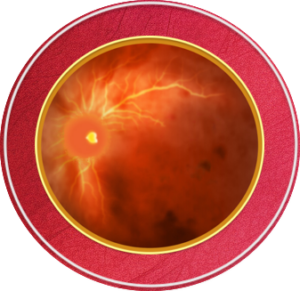
A retinal vein occlusion is a blockage of a blood vessel (vein) that is responsible for taking blood away from the retina. Because blood cannot drain properly from the retina, blood flow backs up, and hemorrhages and areas of swelling may develop. RVO can cause vision loss by provoking swelling of the macula - the central area of the retina (macular edema), bleeding in the retina, or abnormal blood vessel growth (neovascularization) in the eye.
Symptoms may include:
Blurred vision
Sudden loss of vision
Blind spot in vision
Pain or pressure in the eye
Distorted vision
Increased floaters
References
American Society of Retina Specialists (ASRS). Branch retinal vein occlusion. 2020. https://www.asrs.org/patients/retinal-diseases/24/branch-retinal-vein-occlusion.
Cleveland Clinic. Retinal vein occlusion (RVO). 2019. https://my.clevelandclinic.org/health/diseases/14206-retinal-vein-occlusion-rvo.
Flaxel CJ, et al. Retinal vein occlusion Preferred Practice Pattern®. Ophthalmology. 2020;127:P288-P320.
Buehl W, Sacu S, Schimdt-Erfurth U. Retinal Vein Occlusions. Dev Ophthalmol. 2010;46:54-72.
All URLs accessed 3/1/22.
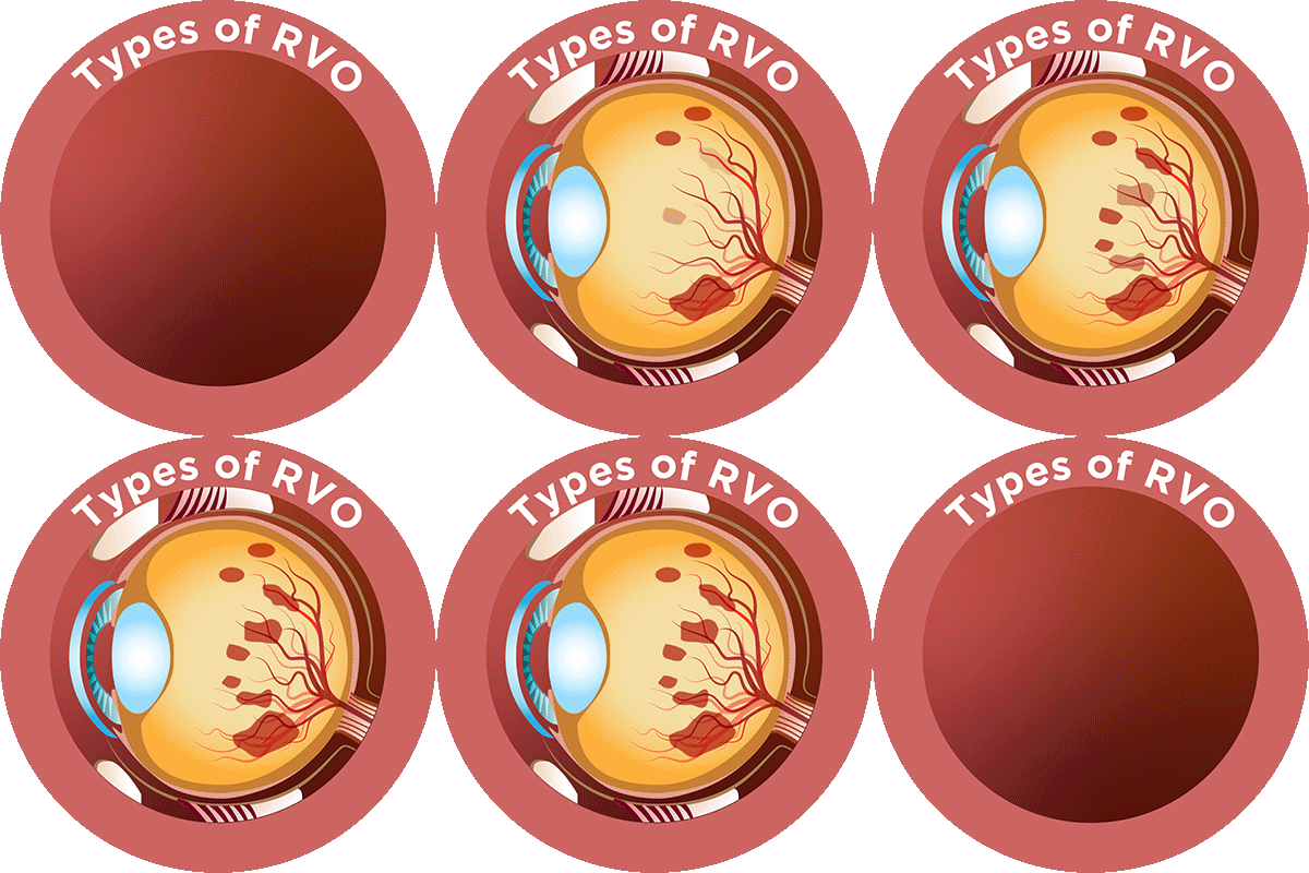
Central retinal vein occlusion occurs when blood flow from the main vein of the retina is blocked. These patients have a higher chance of vision loss and needing treatment than those who have a branch retinal vein occlusion. A branch retinal occlusion is a blockage of one or more of the smaller branches that stem off of the main vein. These branches lead to four different areas of the retina, each draining one quarter of the eye.
References
Morris R. Retinal vein occlusion. Kerala J Ophthalmol. 2016;28:4-13.
Lowth M. Retinal vein occlusion. 2017. https://patient.info/eye-care/visual-problems/retinal-vein-occlusion.
American Society of Retina Specialists (ASRS). Central retinal vein occlusion. 2020. https://www.asrs.org/patients/retinal-diseases/22/central-retinal-vein-occlusion.
All URLs accessed 3/1/22.
Unfortunately, there is no treatment to remove retinal vein blockages. After determining the location and the extent of retinal damage, treatment focuses on controlling complications of RVO as well identifying any risk factors and addressing them.
Macular edema, which is build up of fluid and swelling of the macula located in the center of the eye, is the leading cause of vision loss from a vein occlusion. Optical coherence tomography (OCT) imaging of the retina will help your eye care provider diagnose and monitor your treatments; it is a non-invasive test that uses light waves to take pictures of the layers of the retina. This test will be repeated at your follow-up visits to determine if and when more treatment is necessary.
Injection of an anti-vascular endothelial growth factor (anti-VEGF) agent is the first-line of treatment. Patients usually receive monthly injections for the first 3 months, with additional injections on an as-needed basis. Steroid injections are another treatment option if the swelling is not resolving. Sometimes a laser is needed to seal off leaky vessels.
References
American Society of Retina Specialists (ASRS). Branch retinal vein occlusion. 2020. https://www.asrs.org/patients/retinal-diseases/24/branch-retinal-vein-occlusion.
Cleveland Clinic. Retinal vein occlusion. 2019. https://my.clevelandclinic.org/health/diseases/14206-retinal-vein-occlusion-rvo.
ASRS. Central retinal vein occlusion. 2020. https://www.asrs.org/patients/retinal-diseases/22/central-retinal-vein-occlusion.
WillsEye Hospital. Central retinal vein occlusion (CRVO). https://www.willseye.org/central-retinal-vein-occlusion-crvo/.
All URLs accessed 3/1/22.
- Complete annual physical along with blood work can help detect risk factors.
- Treat and control high blood pressure, high cholesterol, and diabetes.
- Make healthy lifestyle choices including a regular exercise regimen, low fat diet, and quitting smoking.
- Keep up with routine eye exams to monitor your eye pressure for glaucoma, a risk factor for retinal vein occlusion development.10
References
Cleveland Clinic. Retinal vein occlusion. 2019. https://my.clevelandclinic.org/health/diseases/14206-retinal-vein-occlusion-rvo.
WillsEye Hospital. Central retinal vein occlusion (CRVO). https://www.willseye.org/central-retinal-vein-occlusion-crvo/.
Jumper JM, RVO workup: When it’s necessary and what to order. Retinal Specialist. 2015. https://www.retina-specialist.com/article/rvo-workup-when-its-necessary-and-what-to-order.
All URLs accessed 3/1/22.
- Can a retinal vein occlusion be prevented?
- What lifestyle changes will help me prevent another occlusion?
- Do I need to see my primary care doctor for a complete physical and bloodwork?
- What symptoms may I experience?
- Will I need treatment?
- How often will I need to see the specialist?
- Can treatment help recover my vision?
Use this tool for an easy symptom guide and questions to ask your primary care and eye care providers: RVO Checklist
Taking part in a clinical trial can be a great way to help improve treatment for your eye disease. Your participation can help both you and others who may benefit from the treatment if it is approved by the US Food and Drug Administration in the future. Here are a few things to consider about participating in a clinical trial:
- Ask your eye doctor about clinical trials related to your eye disease.
- If your eye doctor says you may be a good candidate for a clinical trial, don’t be afraid to ask questions about the trial. Some possible questions: What would you need to do to participate? How many visits/appointments are needed related to the trial? Is there any assistance with transportation costs for trial-related appointments?
- If you speak another language, find out if trial-related paperwork is available in your native language.
Before you join the trial, leaders of the clinical trial will let you know what you need to do to participate. They can also let you know how you can leave the trial if you choose to do so. Providing information about the trial and letting you know about any potential treatment side effects is called informed consent.1
For more information about clinical trials, click here to access a variety of educational videos and information about medical research, as well as important questions to ask - provided by the US Department of Health and Human Services (HHS).2 Sharing this information with your provider can help you identify appropriate trials for your retinal condition and support discussions on whether or not participation in a clinical trial is right for you.
References
- US Food and Drug Administration. Clinical trial diversity. https://www.fda.gov/consumers/minority-health-and-health-equity/clinical-trial-diversity
- HHS, Office for Human Research Protections. About research participation. https://www.hhs.gov/ohrp/education-and-outreach/about-research-participation/index.html


